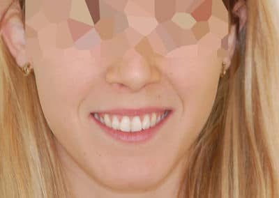
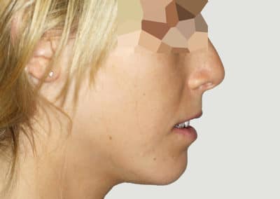
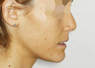
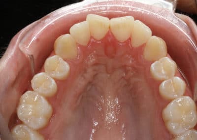
Our smiles:
Orthognathic Surgery

MAKE AN APPOINTMENT
Fatiha B.

A CASE OF A SURGICAL GAP

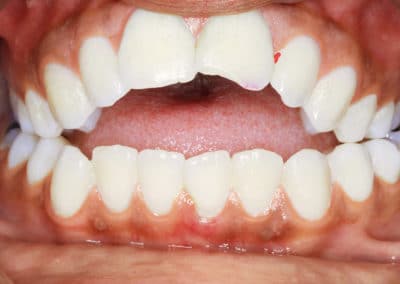
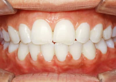
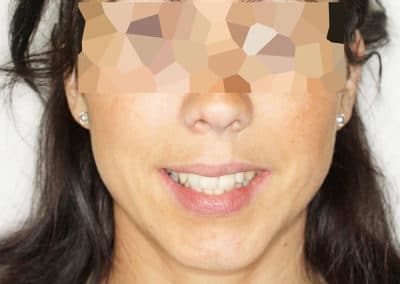
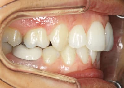
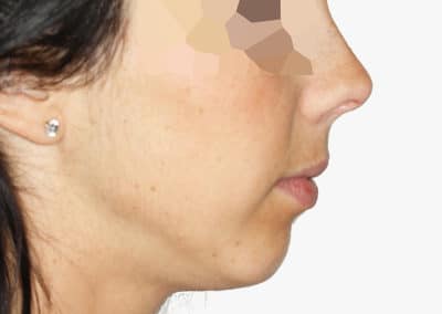
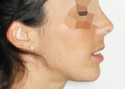
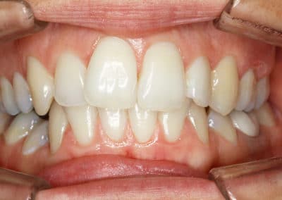
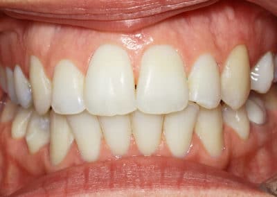
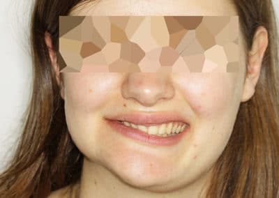
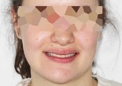
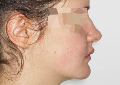
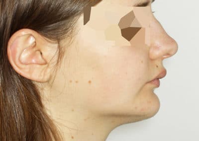
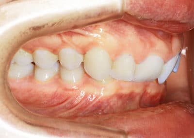
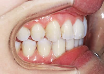
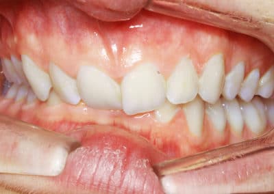
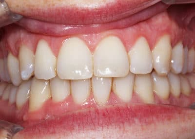
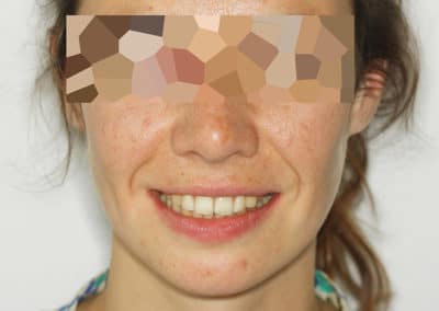
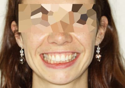
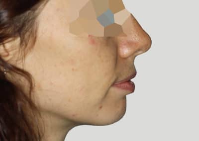
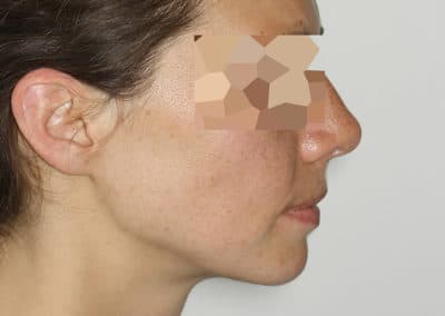
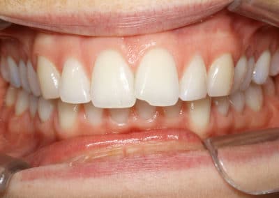
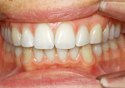
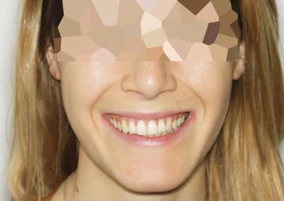
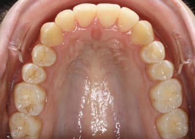
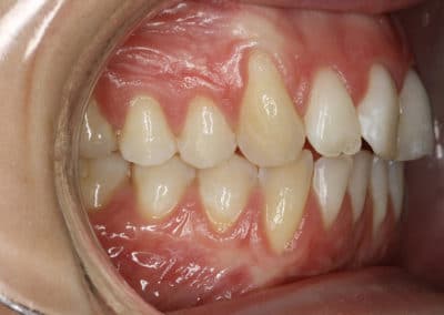
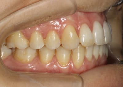
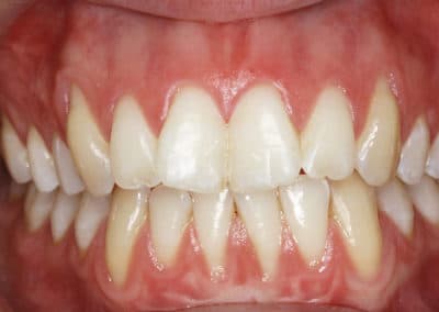
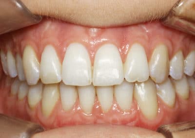
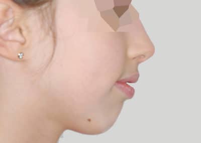
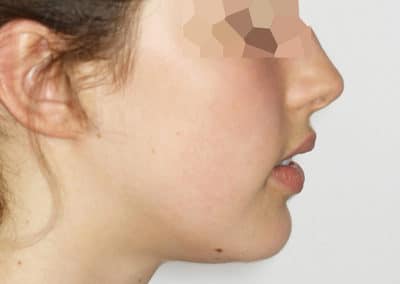
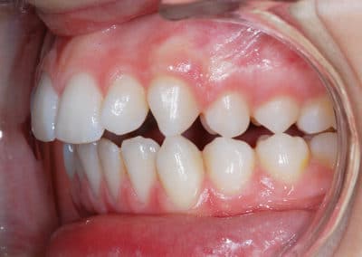
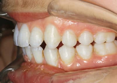
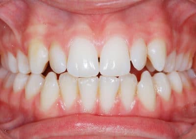
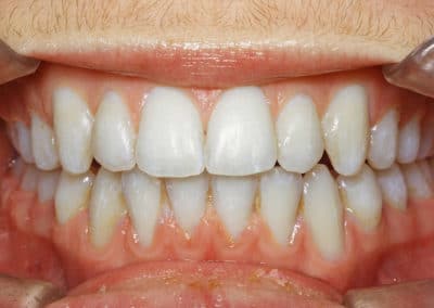
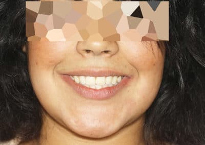
BF presented to an orthodontic consultation with the reason for consultation being her open bite. The clinical and radiological examination revealed a gap related to her skeletal pattern and thumb sucking, a very narrow upper jaw, a very receding chin and lower jaw. On the dental side, there was an inverted smile curve and a 6-mm overhang. The simplified treatment plan was as follows: a surgical maxillary disjunction followed by a mixed lingual treatment, and in a second phase a surgical advancement and counterclockwise rotation of the mandible.

The duration of treatment was 26 months. Rigid wire restraints were placed superiorly and inferiorly from canine to canine on the lingual surfaces. Safety splints were also made.
Emy B.

A CASE OF MAXILLARY MIDLINE DEVIATION

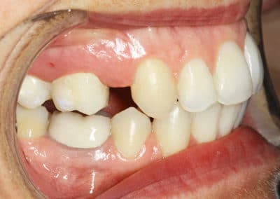
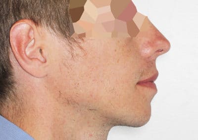
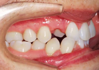
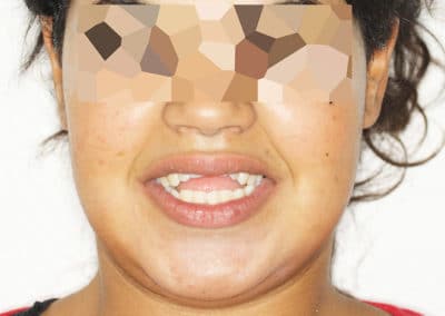
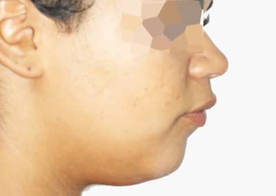
EB presented for consultation because she did not like her left deviated maxillary midline. Clinical and radiological examination revealed the absence of two upper left premolars and one upper right premolar, hence the deviation of her maxillary midline. It also revealed the absence of two mandibular premolars, and the near inclusion of his mandibular premolars. This condition had been generated by a previous treatment. It was also noted that her chin was receding. After showing her a few cases of other patients who had undergone genioplasty and arch symmetrization, the patient decided to follow the following treatment plan: a total lingual treatment combined with genioplasty, with opening of a prosthetic space for the left maxillary premolar #14, thus allowing her maxillary midline to be refocused on her face. On the pictures, you will notice that the space for the premolar has not yet been filled, and that it is undersized. It is not possible to recreate an ideal space, but it is always possible to compensate for this lack of space prosthetically and this is what was planned with her dentist. The patient had been informed before starting the treatment, but we knew that this was not a problem anyway.

The duration of treatment was 24 months. Rigid wire restraints were placed superiorly and inferiorly from canine to canine on the lingual surfaces. Safety splints were also made.
Jade G.

I HAVE A MANDIBULAR ASYMMETRY

JG presented for consultation with mandibular asymmetry and dental misalignment. Clinical and radiological examination revealed a left hypercondylism and a strong mandibular retrogression. The treatment plan was as follows: after confirmation of the hypercondylism with a bone scan, a left condyloscopy was performed. Four months after this procedure, a mixed lingual treatment was performed, and 12 months after the beginning of the treatment, a mandibular advancement surgery for maxillary reaxation and a genioplasty were performed. It should be noted that the symmetry is not perfect because only the basal surgery was performed. For a perfectly symmetrical result, a surgery of the bone contours should have been done in a second step, but as most often the patient was already very satisfied with the improvement obtained.

At the end of the treatment, rigid restraining wires were placed on the lingual surfaces from canine to canine. Safety splints were also made.

Maxime K.

I HAVE A STRONG ASYMMETRY

KM presented to the clinic with a severely deviated lower jaw, a lingual upper right canine and, more generally, all of his overlaps.

The clinical and radiological examination confirmed his reason for consultation and the treatment plan chosen was as follows: a mixed lingual treatment associated with mandibular advancement and maxillary expansion surgery. The patient was informed beforehand that in cases of mandibular asymmetry, there are two components: a basal component that can be corrected by a recentering surgery, and a component that concerns the bony contours that can only be corrected by a contouring surgery, which can only be done in a second step. We also told him that it was rare that contouring surgery still seemed necessary to the patient after basal surgery, because the improvement related to basal surgery is very significant. Also, you will notice on the end photos that a remnant of asymmetry remains. This is indeed the symmetry of the bone contours.

The duration of treatment was 24 months. Rigid wire restraints were placed superiorly and inferiorly from canine to canine on the lingual surfaces. Safety splints were also made.
Mélanie LF.

MY UPPER TEETH ARE TOO FAR FORWARD AND MY JAW IS RECESSED (CLASS II, 1)

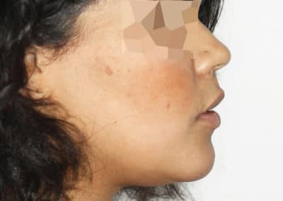
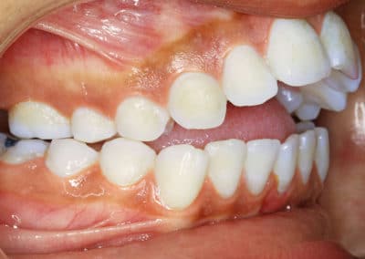
ML presented for consultation with her upper teeth far forward, her lower jaw recessed, and her lower lip hemmed in. The clinical and radiological examination revealed a strong mandibular retrogression, associated with a poorly developed chin button and a large overlap. The simplified treatment plan was as follows: mixed lingual treatment combined with mandibular advancement surgery, plus advancement genioplasty.

After 24 months of treatment, rigid wire restraints were placed superiorly and inferiorly from canine to canine on the lingual surfaces. Safety splints were also made.
Julia O.

I HAVE A GINGIVAL SMILE

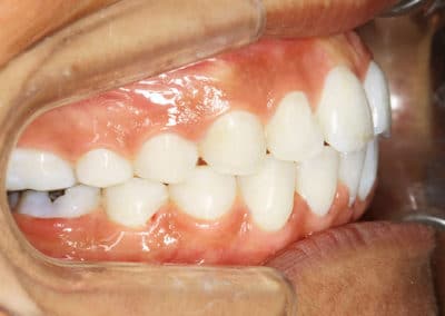
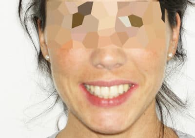
OJ presented for orthodontic consultation with her gummy smile and misalignments. Clinical and radiological examination revealed a vertical overgrowth of her maxilla, slightly more pronounced on the right than on the left, with a deviated mandible on the left and asymmetric functioning of the lip lift muscles. She had no gingival excess, therefore no indication for gingivectomy, nor did she have a short upper lip.

The treatment chosen was therefore a mixed lingual treatment with maxillary impaction and mandibular recentering. Rigid wire restraints were placed superiorly and inferiorly from canine to canine on the lingual surfaces. Safety splints were also made.
Camille P.

I CAN’T BITE ON MY FRONT TEETH

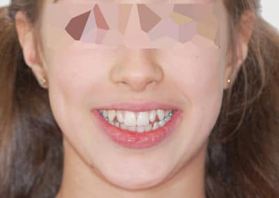
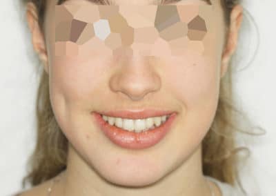
PC came to the orthodontic clinic with a receding chin and the inability to bite on her front teeth.

The diagnosis revealed a 4-mm Class II occlusion, lack of overlap, narrowness of the upper jaw, and a hyperdivergent Class II skeletal pattern. The treatment plan chosen was non-surgical palatal expansion with external – but could have been internal – bands and counterclockwise mandibular advancement surgery, as well as advancement genioplasty during the same surgical time. The treatment lasted 26 months. Rigid wire restraints were placed superiorly and inferiorly from canine to canine on the lingual surfaces. Safety splints were also placed.
Follow me on Instagram